Diagnosis Process
Tests and procedures used to diagnose ACL injuries accurately.
Initial Evaluation
The diagnosis of an ACL injury begins with a detailed patient history. Your healthcare provider will ask about the mechanism of injury, whether you heard or felt a “pop,” the timing of swelling, and any sensation of knee instability. This history often provides valuable clues about the nature and severity of the injury.
Physical Examination
Several clinical tests are used to assess ACL integrity:
Each of the following tests are done manually by hand.
- Lachman test: Considered the most reliable clinical test (sensitivity >90%), it assesses anterior tibial translation. The examiner stabilizes the femur with one hand and pulls the tibia forward with the other while the knee is flexed 20–30 degrees. The examiner is looking for excessive anterior tibial translation or a soft/absent endpoint compared with the other knee, signaling ACL insufficiency. (“anterior” means forward or toward the front of the body. In the knee, anterior tibial translation refers to the shin bone (tibia) moving forward relative to the thigh bone (femur).)
- Anterior drawer test: Evaluates forward movement of the tibia while the knee is bent at 90 degrees. With the patient lying down and the knee bent to 90 degrees, the examiner sits on the foot and pulls the tibia forward with both hands placed behind the upper calf. The examiner is looking for forward tibial movement greater than the unaffected side, especially with a soft endpoint, suggesting ACL laxity or rupture. On the anterior drawer test: Laxity (partial tear or stretched ligament) may show increased forward movement of the tibia with a firm or slightly soft endpoint; the ligament still offers some resistance. Rupture (complete tear) typically shows more pronounced forward movement with a very soft or absent endpoint, meaning there’s little to no resistance from the ACL. The quality of the endpoint, (how abruptly the movement stops), is key to telling the difference.
- Pivot shift test: Assesses rotational stability of the knee. The examiner lifts the leg, applies inward rotation to the tibia, and applies a valgus force while flexing the knee to assess for a clunk indicating subluxation and reduction. A palpable “clunk” as the tibia subluxes and then reduces during flexion, indicating anterolateral rotary instability from an ACL tear. (“subluxes” means partially dislocates; when a joint briefly shifts out of its normal position but then returns. In the pivot shift test, it refers to the tibia sliding slightly out of place (anteriorly and laterally) due to ACL deficiency, then snapping back into alignment as the knee flexes.)
- Examination for associated injuries: Including meniscal tears, other ligament damage, and cartilage injuries. Also done by hand, this includes palpation, specific stress tests (like McMurray for meniscus, valgus/varus stress for MCL/LCL), and observing for joint line tenderness, swelling, or mechanical symptoms.
The McMurray test checks for meniscal tears by rotating the lower leg while extending and flexing the knee. The examiner looks for a click, pop, or pain along the joint line, which may indicate a torn meniscus being caught between the femur and tibia. Valgus stress is when the examiner applies force inward on the knee (from the outside), stressing the medial (inner) structures like the MCL. Varus stress is when force is applied outward on the knee (from the inside), stressing the lateral (outer) structures like the LCL.)
You may hear the term “unhappy triad” which refers to injuries with these structures typically associated together; ACL, MCL and medial meniscus.
Limitations: While these tests are highly effective, their accuracy depends on examiner experience, patient relaxation, and the absence of confounding factors (e.g., muscle guarding, swelling). For more definitive diagnosis, imaging (e.g., MRI) is often used to confirm findings. (At the same time, remember that even modern advanced imaging can be imperfect and not wholly conclusive.)
Imaging Studies
Diagnostic imaging confirms the diagnosis and assesses for associated injuries:
- X-rays: While they cannot visualize ligaments directly, they can identify associated fractures or avulsions
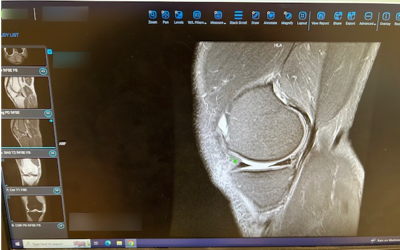
- MRI (Magnetic Resonance Imaging): The gold standard for diagnosing ACL tears, with approximately 95% accuracy. MRI can also reveal meniscal tears, cartilage damage, bone bruises, and other ligament injuries. However, the MRIs don’t necessarily show everything. Or even if they indicate issues, they may not be definitive in terms of degree.
- Ultrasound: Sometimes used for dynamic assessment but less common than MRI
Grading ACL Injuries
ACL injuries are typically categorized as:
- Grade 1: Mild damage; the ligament is stretched but still provides knee stability
- Grade 2: Partial tear of the ligament; rare in ACL injuries
- Grade 3: Complete tear; the ligament is split into two pieces and the knee is unstable
Time
Orthopedists may want to have some time in between injury and imaging studies or after imaging studies and making surgical decisions to see how things progress. Since surgery isn’t likely to happen immediately if there are major swelling issues and such, it’s likely useful to to to physical therapy for awhile and see how things progress. (If there were other associated injuries, those might have to be dealt with earlier of course.) The point is, it might be useful to take some time in between initial consult and more definitive tests to do some “prehab” to see how things develop.
See Also
📰 Web Articles
- Anterior Drawer Test – Cleveland Clinic
- Lachman Test – Physiopedia
- Grades of ACL Tears Explained
- The Pivot Shift Test
🎥 Videos
- Lachman Test | Cruciate Ligament (YouTube)
- Anterior Drawer Test | Anterior Cruciate Ligament Rupture (YouTube)
- Diagnosing ACL Insufficiency: Anterior Drawer and Lachman’s Test Video (YouTube)
- How ACL Injury is Diagnosed and Treated (YouTube)
- How to Read Knee MRI of ACL Tear | Anterior Cruciate Ligament Pain (YouTube)
- ACL Injury ..Diagnosis , Causes , and Treatment Options (YouTube)
- How do you diagnose an ACL RUPTURE?! | Expert Explains Key signs for an ACL tear you need to know! (YouTube)